2012 Alphacoronavirus Detected in Bats in the United Kingdom PDF
Preview 2012 Alphacoronavirus Detected in Bats in the United Kingdom
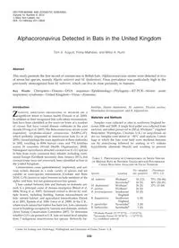
Alphacoronavirus Detected in Bats in the United Kingdom Tom A. August, Fiona Mathews, and Miles A. Nunn Abstract This study presents the first record of coronavirus in British bats. Alphacoronavirus strains were detected in two of seven bat species, namely Myotis nattereri and M. daubentonii. Virus prevalence was particularly high in the previously unrecognized host M. nattereri, which can live in close proximity to humans. Key Words: Chiroptera—Disease—DNA sequence—Epidemiology—Phylogeny—RT-PCR—Severe acute respiratory syndrome—United Kingdom—Virus—Zoonoses. Introduction Z oonotic infections originating in wildlife are a significant threat to human health (Daszak et al. 2000). In addition to their recognized link with rabies transmission, bats have been identified as the reservoir hosts of a number of viruses that have caused disease outbreaks in the past decade (Wong et al. 2007). The Betacoronavirus, severe acute respiratory syndrome-related coronavirus (SARS-CoV), which probably originated in insectivorous bats (Li et al. 2005), caused perhaps the most significant of these outbreaks in 2002, resulting in 8096 human cases and 774 fatalities across 26 countries (World Health Organization, 2004). Subsequent surveillance detected coronavirus (CoV) species in bats from every continent they inhabit, including conti- nental Europe (GenBank taxonomy data, January 2011), but coronaviruses have not previously been identified in bats in the United Kingdom. Coronaviruses cause gastrointestinal, respiratory, and ner- vous system diseases in a wide variety of species and are capable of cross-species transmission (Graham and Baric 2010). CoVs known to infect humans include members of the alpha- and beta- but not gammacoronavirus genera. Close contact between bats and humans or domestic ani- mals is a principal cause of disease emergence (Breed et al. 2006; Wong et al. 2007). Increased contact is driven by mul- tiple factors, including hunting, human encroachment on bats’ natural habitat, and agricultural intensification (Dasak et al. 2001; Epstein et al. 2006; Leroy et al. 2009). In the U.K., where much natural bat habitat has been lost, bats commonly roost in buildings occupied by humans and domestic animals (Joint Nature Conservation Committee 2007). To determine whether CoVs are present in U.K. bats we tested fecal samples from 7 indigenous insectivorous bat species: Barbastella bar- bastellus, Myotis daubentonii, M. nattereri, Plecotus auritus, Rhinolophus ferrumequinum, and R. hipposideros. Materials and Methods Samples were collected at sites in southwest England be- tween 2006 and 2009. A single fecal pellet was collected from each bat, and either preserved in 250 lL RNAlater� (Applied Biosystems, Warrington, Cheshire, U.K.) or snap-frozen on dry ice. Samples were stored at - 80�C until analysis. Cotton bags in which the bats were held were sterilized between use by autoclaving followed by soaking in 6% sodium hypochlorite (domestic bleach) and washing to prevent Table 1. Prevalence of Coronavirus in Seven Species of British Bats by Reverse Transcriptase-Polymerase Chain Reaction Analysis of Fecal Samples Species Location No. sampled (no. positive) Prevalence (95% CI) M. nattereri Wythama 16 (12) 75% (54–96) Savernakeb 16 (9) 56% (32–81) M. daubentonii Wythama 30 (5) 17% (3–30) P. auritus Wythama 26 (0) R. ferrumequinum Southwest England 15 (0) R. hipposideros Southwest England 6 (0) P. pipistrellus Savernakeb 2 (0) B. barbastellus Savernakeb 1 (0) aWytham Woods (51�77’27’’N, - 1�33’41’’E). bSavernake Forest (51�39’96’’N, - 1�67’75’’E). CI, confidence interval. Centre for Ecology and Hydrology, Wallingford, and University of Exeter, Exeter, U.K. VECTOR-BORNE AND ZOONOTIC DISEASES Volume 12, Number 6, 2012 ª Mary Ann Liebert, Inc. DOI: 10.1089/vbz.2011.0829 530 FIG. 1. Neighbor joining phylogeny of representative coronavirus (CoV) RNA-dependent RNA polymerase (RdRP) se- quences (366 bp), including the new strains (in bold type) found in U.K. bats. Bootstrap values (1000 replicates) are indicated as percentages where the value was greater than 70%. CoVs known to infect humans are indicated by a closed circle (�). The common name of hosts is given for CoV not derived from bats. Scale bar indicates base differences per sequence. 531 cross-contamination. Procedures were approved by the Biosciences Ethics Committee, University of Exeter, and car- ried out under the appropriate Natural England license. Fecal pellets stored in RNAlater were homogenized in situ, whereas samples frozen on dry ice were homogenized in 300 lL phosphate-buffered saline (PBS; pH 7.2). Selected fecal homogenates were spiked with 0.2 PFU of human cor- onavirus NL63 stock (grown and titrated in LLC-MK2 cells) to act as positive controls. PBS or RNAlater served as negative controls. RNA was extracted from 100 lL of fecal homogenate using a viral RNA Mini Kit (Qiagen, Crawley, West Sussex, U.K.). The eluted RNA (8 lL of 60 lL) was random primed reverse transcribed (SuperScript II�; Invitrogen, Paisley, U.K.). Semi-nested PCR using ImmoMix� (Bioline, London, U.K.) was used to amplify a *440 bp CoV-specific region of the RNA-dependent RNA polymerase (RdRP). The PCR was performed as previously described (de Souza Luna 2007), except that first round primers were used at 1 lmol/L, and the number of thermocycles was increased to 35 for the second round of amplification. Representative PCR-positive samples were gel purified (QIAquick kit; Qiagen), cloned (pGEM-T vector; Promega, Madison, WI), and sequenced using vector- specific T7 and SP6 primers (BigDye� Terminator vs3.1 Cycle Sequencing Kit and ABI 3730 DNA analyzer; Applied Bio- systems, Carlsbad, CA). All consensus sequences were sub- mitted to GenBank (accession numbers JF440349–JF440366). The novel sequences were aligned with sequences from GenBank using ClustalW in BioEdit 7 (http://www.mbio .ncsu.edu/bioedit/bioedit.html). Phylogenetic analysis was undertaken with MEGA5 (www.megasoftware.net) using a 366-bp region of the RdRP common to all sequences included in the analysis. Attempts to isolate virus in tissue culture (pig kidney epithelial cells) were unsuccessful (data not shown). Statistical analyses of prevalence were performed with R version 2.11.0 (www.r-project.org) Results and Discussion Fecal samples from 112 bats were processed and cor- onavirus RdRP RNA was detected in 2 of the 7 bat species examined (Table 1). Five of 30 M. daubentonii and 21 of 32 M. nattereri samples were positive. All positive and negative controls yielded the expected results. The viral RdRP sequences from the feces of both the British bat species fall into a phylogenetic subclade that originates within the main Alphacoronavirus clade (Fig. 1). The se- quences from European vespertilionid bats form a subclade with 99% bootstrap support. Maximum likelihood (not shown) and neighbor-joining algorithms produced equivalent trees. Some inter-sample sequence variation existed, though clones from a single sample differed by no more than 4 bp. The se- quences from British M. daubentonii are closely related to se- quences obtained from M. daubentonii sampled in Germany (Fig. 1). The sequences from M. nattereri represent the first record of a coronavirus from this bat species and form a novel, well-supported clade (100% bootstrap value). The M. nattereri clade further divides into two groups which correspond to the two sites (47 km apart) at which the species was sampled. Further sampling and analyses of additional alphacoronavirus loci will be needed to interpret this observation. The high prevalence of alphacoronavirus in M. nattereri may be a characteristic of the virus strain. However, unlike M. daubentonii, all M. nattereri samples were collected from maternity roosts, supporting findings from previous studies that have also found high virus prevalence in such roosts (Gloza-Rausch et al. 2008; Drexler et al. 2011). Within these roosts juveniles are immunologically naı¨ve and contact rates are high, which may contribute to the high virus prevalence. The prevalence observed in M. daubentonii is similar to ob- servations from the Netherlands (Reusken et al. 2010). A lo- gistic regression model, including sex, age, location, and species, showed that M. nattereri had a significantly higher prevalence than M. daubentonii ( p = 0.0002), and across both species suggested a trend toward higher prevalence in juve- niles (57% in 21 juveniles, 33% in 43 adults, p = 0.09). The British bat alphacoronavirus strains we identified are distantly related to the zoonotic pathogen SARS-CoV. How- ever, some CoVs are able to switch hosts (Graham and Baric 2010), and evidence suggests that alphacoronaviruses from bats have spilled over to humans in the past (Pfefferle et al. 2009). Therefore, further screening of British bats to better understand CoV epidemiology and to determine whether they harbor other previously unrecognized viruses with zoonotic potential is justified. Acknowledgments We thank Christian Drosten and Petra Herzog for gener- ously donating human coronavirus NL63 and cell line LLC- MK2. We also thank Danielle Linton, Heidi Cooper-Berry, Steven Laurence, and many others who helped collect field samples. Thanks also to William Tyne for assistance with se- quencing, and Stefanie Schafer for reviewing the manuscript. Funding was provided by the Natural Environment Research Council (NERC). Author Disclosure Statement No competing financial interests exist. References Breed AC, Field HE, Epstein JH, et al. Emerging henipaviruses and flying foxes—Conservation and management perspec- tives. Biol Conserv 2006; 131:211–220. Daszak P, Cunningham AA, Hyatt AD. Anthropogenic envi- ronmental change and the emergence of infectious diseases in wildlife. Acta Trop 2001; 78:103–116. Daszak P, Cunningham AA, Hyatt AD. Emerging infectious diseases of wildlife—Threats to biodiversity and human health. Science 2000; 287:443–449. de Souza Luna LK, Heiser V, RegameyN, et al. Generic detection of coronaviruses and differentiation at the prototype strain level by reverse transcription-PCR and nonfluorescent low- density microarray. J Clin Microbiol 2007; 45:1049–1052. Drexler JF, Corman VM, Wegner T, et al. Amplification of emerging viruses in a bat colony. Emerg Infect Dis 2011; 17:449–456. Epstein JH, Field HE, Ludby S, et al. Nipah virus: Impact, origins, and causes of emergence. Curr Infect Dis Rep 2006; 8:59–65. Gloza-Rausch F, Ipsen A, Seebens A, et al. Detection and prev- alence patterns of group I coronaviruses in bats, northern Germany. Emerg Infect Dis 2008; 14:626–631. Graham RL, Baric RS. Recombination, reservoirs, and the modular spike: mechanisms of coronavirus cross-species transmission. J Virol 2010; 84:3134–3146. 532 AUGUST ET AL. Joint Nature Conservation Committee. Second Report by the UK under Article 17 on the implementation of the Habitats Di- rective from January 2001 to December 2006. Peterborough, JNCC, 2007. Leroy EM, Epelboin A, Mondonge V, et al. Human Ebola out- break resulting from direct exposure to fruit bats in Luebo, Democratic Republic of Congo, 2007. Vector-Borne Zoonotic Dis 2009; 9:723–728. Li WD, Shi ZL, Yu M, et al. Bats are natural reservoirs of SARS- like coronaviruses. Science 2005; 310:676–679. Pfefferle S, Oppong S, Drexler JF, et al. Distant relatives of severe acute respiratory syndrome coronavirus and close relatives of human coronavirus 229E in bats, Ghana. Emerg Infect Dis 2009; 15:1377–1384 Reusken CBEM, Lina PHC, Pielaat A, etal. Circulation of group 2 coronaviruses in a bat species common to urban areas in Western Europe. Vector-Borne Zoonotic Dis 2010; 10:785–791. Wong S, Lau S, Woo P, et al. Bats as a continuing source of emerging infections in humans. Rev Med Virol 2007; 17:67–91. World Health Organization. Summary of probable SARS cases with onset of illness from 1 November 2002 to 31 July 2003. Available at http://www.who.int/csr/sars/country/ table2004_04_21/en/index.html, accessed July, 2011. Address correspondence to: Miles Nunn Centre for Ecology and Hydrology Maclean Building Benson Lane Crowmarsh Gifford Wallingford, Oxfordshire, OX10 8BB United Kingdom E-mail:
