1 CARDIOLOGY FOR PRIMARY CARE Lois E Brenneman, MSN, ANP, FNP, C Anterior Heart ... PDF
Preview 1 CARDIOLOGY FOR PRIMARY CARE Lois E Brenneman, MSN, ANP, FNP, C Anterior Heart ...
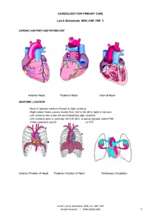
CARDIOLOGY FOR PRIMARY CARE Lois E Brenneman, MSN, ANP, FNP, C CARDIAC ANATOMY AND PHYSIOLOGY Anterior Heart Posterior Heart Internal Heart ANATOMIC LOCATION - Most of anterior surface of heart is right ventricle - Right atrium forms narrow border from 3rd to 5th rib to right of sternum - Left ventricle lies to the left and behind the right ventricle. - Left ventricle apex is normally 5th ICS-MCL w apical impulse called PMI. - Other chambers not ID on P/E Anterior Position of Heart Posterior Position of Heart Pulmonary Circulation © 2001 Lois E. Brenneman, MSN, CS, ANP, FNP - all rights reserved www.npceu.com 1 CARDIACLANDMARKS - 2nd ICS to right and left of sternum called BASE . - Left atrium: most posterior portion of heart; when enlarged, extends posteriorly and to the right . - Tip of left ventricle is called APEX (5th ICS-MCL) When left ventricle enlarges -> it extends laterally and downward hence PMI will no longer be in 5th ICS-MCL but will be displaced (e.g CHF). AUSCULTATION SITES - Aortic: 2nd ICS,right sternal border (2ICS-RSB) - Pulmonic: 2nd ICS,left sternal border (2ICS-LSB) - Tricuspid: Left lower sternal border (LLSB) - Mitral: Cardiac apex - Erb's point: 3rd ICS os frequently the area to which pulmonic or aortic sounds radiate. Four classic auscultatory areas correspond to points over the precordium at which events originating in each valve are best heard. - Areas do not necessarily related to anatomic position of the valve - Sounds heard in the area not necessarily directly produced by the valve that names the area. NEURAL STIMULATION OF THE HEART - Sympathetic stimulation (norepinephrine) produces marked increase in HR and contractility - Parasympathetic stimulation (acetylcholine) slows the heart (Vagus). - Several receptors that provide circulatory info to medullary cardiovascular center in brain - Cardio-excitatory and cardioinhibitory areas that regulate neural output to sympathetic and parasympathetic fibers. © 2001 Lois E. Brenneman, MSN, CS, ANP, FNP - all rights reserved www.npceu.com 2 CARDIAC VALVES A-V valves: between atria and ventricles (1st sound) Tricuspid (3 leaflets): right Mitral (2 leaflets): left Semilunar valves: between atria and arteries(2nd sound) Aortic: aorta and L ventricle Pulmonic: pulmonary arteryand R ventricle Semilunar Valve Atrioventricular Valves Valvular Stenosis Heart Valves COMPONENTS OF CARDIAC CYCLE Isovolumetric contraction: Time between closure of AV valve and opening of semilunar valve. When pressure in RV exceeds diastolic press in PA: pulmonic valve opens If stenotic hear pulmonic ejection click. When pressure in left ventricle exceeds the diastolic pressure in aorta: aortic valve opens. If stenotic will hear an ejection click. Systolic periodof ejection: time between opening and closing of semilunar valves Point at which ejection is completed and the aortic and left ventricular curves separate is called the incisura or dicrotic notch, and is simultaneous with the aortic component of S2 or closure of the aortic valve. (commonly written A2). © 2001 Lois E. Brenneman, MSN, CS, ANP, FNP - all rights reserved www.npceu.com 3 Pulmonic valve closes at the point when right ventricular pressure falls below the pulmonary diastolic pressure and is the pulmonic component of S2 (commonly written P2). Isovolumetric relaxation: time between closure of semilunar valves and opening of atrioventricular valves. Tricuspid valve opens when pressure in right atrium exceeds right ventricular pressure. Opening snap if tricuspid is stenotic. Mitral valve opens when pressure in left atrium exceeds left ventricular pressure. Mitral opening snap if mitral valve is stenotic. Period of rapid filling of ventricles: Occurs with opening of A-V valves; approximately 80% of ventricularfilling occurs at this time. Third heart sound (S3) may be heard at end of this rapid filling period. Ventricularfilling. Fourth heart sound (S4) may e heard at end of diastole during period of Atrial contraction HEART SOUNDS - Normally only the closing of the heart valves can be heard. - 1st heart sound: closing of A-V valves (mitral, tricuspid) - 2nd heart sound: closing of semilunar valves (aortic, pulmonic). - Opening of valves can be heard only if they aredamaged. - A-V valve (mitral or tricuspid) narrowed or stenotic:opening may be heard as an opening snap (DIASTOLE). Opening snap: refers to opening of a pathologically damaged A-V valve (mitralor tricuspid) that occurs duringdiastole. - Semilunar valve (aortic or pulmonic) stenosis: opening may be heard as an ejection click (SYSTOLE). Ejection click: refers to the opening of a damaged semilunar valve that occurs during systole. - Sequence of valves opening and closing: Mitral valve close; tricuspid valve close (1st heart sound) Pulmonic valve open; Aortic valve open Aortic valve close; pulmonic vale close (2nd heart sound) Tricuspid valve open; mitralvalve open - S1 (1st heart sound): closing of A-V valves (mitral, tricuspid) © 2001 Lois E. Brenneman, MSN, CS, ANP, FNP - all rights reserved www.npceu.com 4 - First heart sound heard loudest at APEX - Splitting of first heart sound may be heard in tricuspid area. - S2 (2nd heart sound): closing of semilunar valves (aortic, pulmonic) - Heard loudest at base. - A2 and P2 indicate the aortic and pulmonic component of S2, respectively. A2 normally precedes P2 (aortic valve closes before pulmonic valve) - With inspiration, intrathoracic pressure lowers thus drawing more blood from superior and inferior venae cavae into right hear thus right ventricle enlarges and takes longer for all of blood to be ejected into PA. - Accordingly, pulmonic valve stays open longer and P2 occurs later in inspiration compared w expiration causing aphysiologic split of s2 - S3 (3rd heart sound) Period of rapid filling of ventricles: - Normal only in children and young adults - Occurs w opening of A-V valves (during period of rapid filling of ventricles) - approximately 80% of ventricularfilling occurs at this time. - May be heard at end of this rapid filling period. - It occurs 120-170 msec after S2; period same as it takes to say the "me" in "me too"; "me" = S2; "too" = S3. - S3 normal in children and young adults; when present in individuals >30, signifies volume overload to ventricle which could be secondary to valvular lesions and CHF - S4 (4th heart sound): Atrial contraction (atrial kick) - Normal only in children and young adults - Occurs at end of diastole and is responsible for additional 20% of ventricularfilling. - May be heard at end of diastole during period of Atrial contraction - Normal in children and young adults. When present in individuals over the age of 30, it indicates a noncompliant, or "stiff" ventricle. (e.g. HTN) - The interval from S4 to S1 is approximately the time it takes to say "middle." The "mi" is the S4 whereas the "ddle" is the S1. Note that "mi" is much softer than "ddle," quit similar to the S4-S1 cadence. - Pressure overload on a ventricle causes concentric hypertrophy, which produces a non- compliant ventricle. - CAD is a major cause of stiff ventricle. - Gallop sounds or rhythms: Presence of an S3 or S4 creates a cadence resembling the gallop of a horse called gallop sounds or rhythms. © 2001 Lois E. Brenneman, MSN, CS, ANP, FNP - all rights reserved www.npceu.com 5 MURMURS GENERAL CONCEPTS - Valves can only do two things: OPEN AND CLOSE. - Normally open and close noiselessly - MURMURS occur when there is valve malfunction -> turbulent blood flow through a valve - Most murmurs involvemitral and/or aortic valve - Pulmonic and tricuspid valve murmurs are not common - We can surmise what is going on the basis of when we hear the murmur (systole or diastole) and where we hear the murmur (apex versus base). - Diligent auscultatory characterization of heart murmurs and ancillaryphysical findings often provide adequate basis for dx STENOSIS: failure of valve to open completely Significant problems when valve opening is reduced to ½ of normal Degree of stenosis estimated by development of abnormal pressure gradient across valve Stenosis results in extra pressure work for heart Blood must be forced thru high resistance of narrow opening - Generally progresses slowly over years to decade allowing heart to compensate - Compensationvia dilation and hypertrophy Aortic Stenosis: Etiology: - Post-inflammatory valvular scarring due to rheumatic heart disease (common) - Valvular calcificationwith aging (common) - Congenital anomaly: bivalvewhich is more susceptible than normal trivalve REGURGITATION OR INSUFFICIENCY: inability of valve to close completely - Allowing blood to flow in reverse direction - May occur due to pathologic changes of valve or changes in supporting structures around valve - May develop suddenly due to valvular infection or rupture of supporting papillary muscle. - Sudden regurgitation is poorly tolerated due to no compensation - Results in extra volume work for heart as more blood must be pumped to maintain adequate forward flow Diseased valves may have both stenosis and regurgitation: one problem usually predominates © 2001 Lois E. Brenneman, MSN, CS, ANP, FNP - all rights reserved www.npceu.com 6 SYSTOLIC MURMURS: - Midsystolic ejection murmurs: produced by forward flow through the aortic and pulmonic valves - Pansystolic regurgitant murmurs Produced by backflow through the atrioventricular valves or flow from left to right ventricle in a VSD DIASTOLIC MURMURS - Early diastolic: startw second heart sound - Mid-diastolic: short pause after second sound - Late diastolic or presystolic: due to atrial contraction DESCRIPTION-GRADING OF MURMURS - describe murmur: timing, radiation and point of intensity - grade murmur: auscultatory characteristics - differentiate: systolic vs diastolic DESCRIBE MURMUR : Timing with respect to cardiac cycle, location, radiation, duration, intensity, pitch, quality, relationship to respiration, relationship to body position . Timing as to diastole and systole is paramount. Systolic murmur begin w or after S1? end before or after S2? Entire systolic period (holosystolic, pansystolic)? Systolic ejection murmur (begins after S1 and ends before S2)? early-mid-late systolic murmur? Holodiastolic: throughout diastole. Best heard? Radiation: axilla? neck? back? GRADE MURMUR: Intensity I: Low intensity; often not heard by inexperienced II: Low intensity, usually audible by inexperienced III: Medium intensity without a thrill. IV: Medium intensity with a thrill. V: Loudest murmur that is audible when stethoscope on chest; associated with a thrill. VI: Loudest intensity; audible when stethoscope is removed from chest; associated w a thrill In general, intensity of a murmur tells you nothing about the severity of the clinical state! Quality: rumbling, blowing, harsh, musical, machinery, scratchy. © 2001 Lois E. Brenneman, MSN, CS, ANP, FNP - all rights reserved www.npceu.com 7 CARDIAC RHYTHM AND RATE - Sinus Arrhythmia: increase in heartrate w inspiration. - NOT TRUE ARRHYTHMIA but physiologic response to decrease in left ventricular volume during inspiration. blood in right ventricle is pumped into large capacitance bed of lungs therefore return of blood from lungs to heart is decreased and left atrium and left ventricle become smaller. - Atrial receptors trigger a reflex tachycardia that compensates for the decreased left ventricular volume aka sinus arrhythmia. - Timing the cardiac cycle: needed to interpret heart sounds - To interpret heart sounds accurately, must time S1-S2 - Most reliable way of ID S1 and S2 is to palpate carotid artery. - Use right hand to position stethoscope and left hand to palpate carotid artery - The sound that preceded carotid pulse is S1; S2 follows pulse. - Do not use radial pulse as time delay from S1 to radial pulse is significant and will result in errors in timing. CARDIAC ARRHYTHMIAS Rhythmicity (automaticity): intermittent spontaneous generation of action potentials Rate determined by relative influx of Na+ and Ca++ vs efflux of K+ Cell with the fastest rate of spontaneous depolarization becomes become pacemaker for rest of heart - SA node in the normal heart (in right atrium) Other myocardial cells capable of becoming pacemaker (spontaneous depolarization) in certain circumstances (usually abnormal i.e. arrhythmias) Rhythmicity influenced by drugs, electrolyte balance and autonomic nervous system NORMAL CONDUCTION/CONTRACTION SEQUENCE Conduction System: SA node -> spread contiguouslycell to cell in atria -> bundle branches in atria carry impulse more rapidly then other atrial cells -> av node * -> atrial contraction(when atria have depolarized) -> slowing of impulse occurs after AV node ->Purkinje cells (fibers) where impulse travels downintraventricular septumtoward apex -> divides into right and left bundle branches which travel down left and right side of intraventricular septum - > penetrate ventricular muscle mass from endocardial side-> spread contiguouslycell to cell through ventricle (toward epicardial surface) -> ventricular contraction * at AV junction located in posterior septal wall of right atrium just behind ventricle Note: fibrous skeleton which separates atria from ventricle thus prevents direct transmission of impulse from atria to contiguous ventricular cells © 2001 Lois E. Brenneman, MSN, CS, ANP, FNP - all rights reserved www.npceu.com 8 P wave: Atrial depolarization P-R interval: Atrial, AV node and Purkinje depolarization Q wave: Septal depolarization R wave: Apical depolarization S wave: Depolarization of lateral walls (base) T wave: Ventricular repolarization SEQUELAE OF CARDIAC ARRHYTHMIAS - Sudden death - Syncope - Heart failure - Dizziness - Palpitations - Asymptomatic Bradycardia (bradyarrhythmias) : heart rate < 60 bpm Tachycardia (tachyarrhythmias): heart rate > 100 bpm -Supraventricular tachycardia: arise from atria or AV junction - Ventricular tachycardia: arise from ventricles Ventricular arrhythmias tend to be more symptomatic (can be life-threatening) MECHANISM OF ARRHYTHMIA - Alteration of automaticity - originate from single cell or abnormal interactions between cells - can result in bradycardia or tachycardia - Accelerated automaticity - increasing rate of depolarization or changing threshold potential - sinus tachycardias, escape rhythms and accelerated AV nodal rhythms - Triggered tachycardia: oscillations of transmembrane potential at end of AP - oscillations reach threshold and produce arrhythmia - can be exaggerated via pacing or catecholamines/drugs (digoxin toxicity-> atrial tach) - Re-entry (or circus movement): results in tachycardia - wave of depolarization travels in 1 direction around a ring of cardiac tissue - circus movement results where time to conduct around ring longer than recovery of any tissue within ring - Accounts for majority of paroxysmal tachycardias NORMAL RHYTHM - Sinus Rhythm or NSR © 2001 Lois E. Brenneman, MSN, CS, ANP, FNP - all rights reserved www.npceu.com 9 ABNORMAL RHYTHMS SINUS RHYTHMS: P waves upright in leads I and II of EKG; inverted in AVR and V1 - Sinus Arrhythmia: normal variation in children and young adults - Fluctuations in autonomic tone -> phasic changes in sinus discharge rate - Inspiration: parasympathetic tone falls -> HR increases; expiration -> HR falls - Sinus Bradycardia: less than 60 bpm day or 56 bpm night - Usually asymptomatic unless very slow - Normal in athletes and elderly - Etiology - Hypothermia, hypothyroidism, cholestatic jaundice, raised IC - Beta blockers, digitalis, antiarrhythmic drugs - Acute ischemia and infarction of sinus node - chronic degenerative changes (fibrosisatrium, sinus node) - TREATMENT: - Acute symptomatic: atropine 600 ug - Persistent irreversible: permanent pacemaker - Sinus Tachycardia: greater than 100 bpm - Etiology: Fever, exercise, emotion, pregnancy, anemia, cardiac failure wi compensatory sinus tachycardia, thyrotoxicosis, catecholamine excess, primary sinus tachycardia (rare) - TX: correct underlying cause; use of beta-blockers PATHOLOGICAL BRADYCARDIAS - Sick Sinus Syndrome (sick node disease): long intervalsbetween p waves on EKG (> 2s) Sinoatrial exit block: pause is exact multiple of basic sinus interval Sinus arrest: intervalis not multiple of basic sinus interval Sinus pauses allow tachyarrhythmias to emerge Tachybrady syndrome: combo of fast and slow supraventricular rhythms TREATMENT : - Permanent pacing w additional antiarrhythmic drugs (tachycardia) - Anticoagulate (thromboembolism common) unless contraindication © 2001 Lois E. Brenneman, MSN, CS, ANP, FNP - all rights reserved www.npceu.com 10
Description: