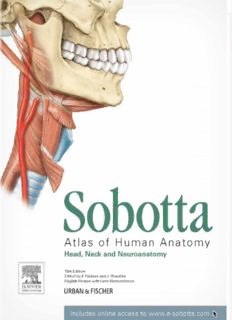
Sobotta Atlas of Anatomy: Head, Neck and Neuroanatomy PDF
Preview Sobotta Atlas of Anatomy: Head, Neck and Neuroanatomy
Sobotta Atl as of Human A n a t o m y Head, Neck and Neuroanatomy 15th Edition Edited by f Paulsen and J Waschko English Version with Latin Nomenclature ELSEVIER URBAN & FISCHER I HtU>A Mtt ittft User's Guide to the Book Introductory pages: • Bulleted lists in figure captions as well as in tables help structu • The introductory pages provide all relevant anatomical informa ring complex facts and provide a better overview. tions concerning the subject of the chapter. Important details • Figures, tables, and text boxes are interconnected by cross- and connections are explained easily to understand. references. • The Dissection Link for each chapter comprises brief and con • Cross-references link the figures to the separate Table Booklet cise tips essential for the dissection of the respective body re with tables of muscles, joints, and nerves, thus providing a suf gion. ficient anatomical knowledge for the exam. • Exam Check Lists provide all keywords for possible exam • Clinical Remarks boxes provide clinical background knowledge questions. concerning the anatomical structures illustrated on the page. • The dissection link on the page indicates if a tip for dissecting Atlas pages: the illustrated anatomical region is available on www.e-sobotta. • The menu bar on top indicates the topics of each chapter, the com. bold print shows the subject of the respective pages. • Important anatomical structures in the figures are highlighted in Appendix: bold print. • List of abbreviations, general terms of direction and position • Small supplement sketches located next to complex views can be found at the end of the book. show visual angles and intersecting planes and, thus, facilitate orientation. • Detailed figure captions explain the relationships of anatomical structures. Perfect Orientation - the New Navigation System The menu bar with the The subject of this page terms printed in bold indicates the subject of the current page. Sketches facilitate orientation in complex figures by showing visual angles and intersecting planes. Important anatomical structures are printed in bold. Figure captions explain anatomical connections The Clinical Remarks concerning the boxes describe medical illustrated structures. contexts to the anatomical structures illustrated on the page. Mostly, these clinical aspects are also of high For pages with this relevance for the exam. dissection link detailed dissection tips can be found on www.e-sobotta. com. The following contents can be found in the other two volumes: 1 General Anatomy m e Orientation on the Body Surface Anatomy -+ Development -► Muskuloskeletal t -* s y System -► Vessels and Nerves Imaging Techniques Integumentary System S l a l e k s o 2 Trunk l u c Surface Anatomy -► Development -► Skeleton -► Imaging -► Muscles -♦ s u M Vessels and Nerves -» Topography, Back Female Breast -♦ Topography, Abdomen and Abdominal Wall d n a y m 3 Upper Extremity o t a Surface Anatomy -► Development -*• Skeleton -► Imaging -► Muscles -► n A Topography -► Sections al r e n e G 4 Lower Extremity 1 Surface Anatomy -► Skeleton -» Imaging Muscles -► Topography ol. Sections V 5 Viscera of the Thorax Heart -» Lungs Oesophagus Thymus -► Topography -» Sections 6 Viscera of the Abdomen s n a Development -► Stomach -► Intestines -► Liver and Gallbladder g Or Pancreas -» Spleen -» Topography -» Sections al n r e t n I 7 Pelvis and Retroperitoneal Space 2 Kidney and Adrenal Gland Efferent Urinary System -► Genitalia ol. Rectum and Anal Canal -► Topography -► Sections V Paulsen, Waschke Sobotta Atlas of Human Anatomy Latin Nomenclature Head, Neck, and Neuroanatomy Translated by T. Klonisch and S. Hombach-Klonisch Sobotta Atlas of Human Anatomy Head, Neck, and Neuroanatomy 15th edition Edited by F. Paulsen and J. Waschke Translated by T. Klonisch and S. Hombach-Klonisch, Winnipeg, Canada 569 Coloured Plates with 627 Figures URBAN & FISCHER München Editors Prof. Dr. Friedrich Paulsen Prof. Dr. Jens Waschke Dissecting Courses for Students More Clinical Relevance in Teaching In his teaching, Friedrich Paulsen puts great emphasis on the fact From March 2011 on, Professor Jens Waschke is Chairman of that students can actually dissect on cadavers of body donors. "The Department I at the Institute of Anatomy and Cell Biology at the hands-on experience in dissection is extremely important not only Ludwig-Maximilians-Universitat (LMU) Munich. " For me, teaching at the for the three-dimensional understanding of anatomy and as the basis department of vegetative anatomy, which is responsible for the for virtually every medical profession, but for many students also dissection courses of both Munich's large universities LMU andTU, clearly addresses the issue of death and dying for the first time. The emphasizes the importance of teaching anatomy with clear clinical members of the dissection team not only study anatomy but also relevance", says Jens Waschke. learn to deal with this special issue. At no other time medical "The clinical aspects in the Atlas introduce students to anatomy in the students will have such a close contact to their classmates and first semesters. At the same time, it indicates the importance of this teachers again." subject for future clinical practice, as understanding human anatomy "The dissection links in the atlas lead to online images that are means more than just memorization of structures." relevant for the dissection. You can print them and take them along. The offered dissection tips are not instructions, but make sure that Professor Jens Waschke (born in 1974) habilitated in 2007 after you are oriented exceptionally well and not 'cutting in the dark'." graduation from Medical School and completing a doctoral thesis at the University of Wuerzburg. From 2003 to 2004 he joined Professor Professor Friedrich Paulsen (born 1965 in Kiel) passed the 'Abitur' in Fitz-Roy Curry at the University of California in Davis for a nine months Brunswick and trained successfully as a nurse. After studying human research visit. Starting in June 2008, he became the Chairman at the medicine in Kiel, he became scientific associate at the Institute of Institute of Anatomy and Cell Biology III at the University of Anatomy, Department of Oral and Maxillofacial Surgery and the Wuerzburg. In 2005, together with his colleagues, Professor Waschke Department of Otolaryngology, Flead and Neck Surgery of the was awarded the Albert Koelliker Teaching Award of the Faculty of Christian-Albrechts-Universität Kiel. In 2002, together with his Medicine in Wuerzburg. In 2006, he was awarded the Wolfgang colleagues, he was awarded the Teaching Award for outstanding Bargmann Prize of the Anatomical Society. teaching in the field of anatomy at the Medical Faculty of the University of Kiel. On several occasions he gained work experience His main research area concerns cellular mechanisms that control the abroad in the academic section of the Department of Ophthalmology, adhesion between cells and the cellular junctions establishing the University of Bristol, UK, where he did research for several months. outer and inner barriers of the human body. The attention is focused on the regulations of the endothelial barrier in inflammation and the From 2004 to 2010 as a University Professor, he was head of the mechanisms, which lead to the formation of fatal dermal blisters in Macroscopic Anatomy and Prosector Section at the Department of pemphigus, an autoimmune disease. The goal is to gain a better Anatomy and Cell Biology of the Martin-Luther-Universität Halle- understanding of cell adhesion as a basis for the development of new Wittenberg. Starting in April 2010, Professor Paulsen became the therapeutic strategies. Chairman at the Institute of Anatomy II of the Friedrich-Alexander- Universität Erlangen. Since 2006, Professor Paulsen is a board member of the Anatomical Society and 2009 he was elected the general secretary of the International Federation of Associations of Anatomy (IFAA). His main research area concerns the innate immune system. Topics of special interest are antimicrobial peptides, trefoil factor peptides, surfactant proteins, mucins, corneal wound healing, as well as stem cells of the lacrimal gland and diseases such as eye infections, dry eye, or osteoarthritis. All business correspondence should be made with: Elsevier GmbH, Urban & Fischer Verlag, Hackerbrucke 6, 80335 Munich, Germany, mail to: [email protected] Addresses of the editors: This atlas was founded by Johannes Sobotta t, former Professor of Professor Dr. med. Friedrich Paulsen Anatomy and Director of the Anatomical Institute of the University in Institut für Anatomie II (Vorstand) Bonn, Germany. Universität Erlangen-Nürnberg German editions: Universitätsstraße 19 91054 Erlangen 1st edition: 1904-1907 J. F. Lehmanns Verlag, Munich Germany 2nd—11th edition: 1913-1944 J. F. Lehmanns Verlag, Munich 12th edition: 1948 and following editions Urban & Schwarzenberg, Munich Professor Dr. med. Jens Waschke 13th edition: 1953, editor H. Becher Institut für Anatomie 14th edition: 1956, editor H. Becher Ludwig-Maximilians-Universität 15th edition: 1957, editor H. Becher Pettenkoferstraße 11 16th edition: 1967, editor H. Becher 80333 München Germany 17th edition: 1972, editors H. Fernerand J. Staubesand 18th edition: 1982, editors H. Fernerand J. Staubesand Addresses of the translators: 19th edition: 1988, editor J. Staubesand Professor Dr. med. Sabine Flombach-Klonisch 20th edition: 1993, editors R. Putz and R. Pabst Urban & Schwarzenberg, Munich Professor Dr. med. Thomas Klonisch Faculty of Medicine 21st edition: 2000, editors R. Putz and R. Pabst Urban & Fischer, Munich Department of Human Anatomy and Cell Science 22nd edition: 2006, editors R. Putz and R. Pabst University of Manitoba Urban & Fischer, Munich 745 Bannatyne Avenue Winnipeg Manitoba R3E 0J9 23rd edition: 2010, editors F. Paulsen and J. Waschke Elsevier, Munich Canada Foreign editions: Bibliographic information published by the Deutsche Nationalbibliothek Arabic edition Modern Technical Center, Damaskus The Deutsche Nationalbibliothek lists this publication in the Deutsche Chinese edition (complex characters) Nationalbibliografie; detailed bibliographic data are available in the Ho-Chi Book Publishing Co, Taiwan Internet at http://www.d-nb.de. Chinese edition (simplified Chinese edition) Elsevier, Health Sciences Asia, Singapore All rights reserved Croatian edition 15th Edition 2011 © Elsevier GmbH, Munich Naklada Slap, Jastrebarsko Czech edition Urban & Fischer Verlag is an imprint of Elsevier GmbH. Grada Publishing, Prague Dutch edition 11 12 13 14 15 5 4 3 2 1 Bohn Stafleu van Loghum, Houten English edition (with nomenclature in English) For copyright concerning the pictorial material see picture credits. Elsevier Inc., Philadelphia English edition (with nomenclature in Latin) All rights, including translation, are reserved. No part of this publication Elsevier GmbH, Urban & Fischer may be reproduced, stored in a retrieval system, or transmitted in any French edition other form or by any means, electronic, mechanical, photocopying, recording, or otherwise without the prior written permission of the Tec & Doc Lavoisier, Paris Greek edition (with nomenclature in Greek) publisher. Maria G. Parissianos, Athen Greek edition (with nomenclature in Latin) Acquisition editor: Alexandra Frntic, Munich Development editor: Dr. Andrea Beilmann, Munich Maria G. Parissianos, Athen Hungarian edition Editing: Ulrike Kriegel, buchundmehr, Munich Production manager: Sibylle Hartl, Munich; Renate Hausdorf, Medicina Publishing, Budapest Indonesian edition buchundmehr, Gräfelfing Penerbit Buku Kedokteran EGC, Jakarta Composed by: Mitterweger & Partner, Plankstadt Italian edition Printed and bound by: Firmengruppe appl, Wemding Elsevier Masson STL, Milan Illustrators: Dr. Katja Dalkowski, Buckenhof; Sonja Klebe, Aying- Japanese edition Großhelfendorf; Jörg Mair, Munich; Stephan Winkler, Munich Igaku Shoin Ltd., Tokyo Cover illustration: Nicola Neubauer, Puchheim Korean edition Cover design: SpieszDesign, Neu-Ulm Elsevier Korea LLC Printed on 115g Quadro Silk Polish edition Elsevier Urban & Partner, Wroclaw ISBN 978-0-7234-3733-8 Portuguese edition (with nomenclature in English) Editora Guanabara Koogan, Rio de Janeiro Portuguese edition (with nomenclature in Latin) Editora Guanabara Koogan, Rio de Janeiro Russian edition Reed Elsevier LLC, Moscow Spanish edition Editorial Medica Panamericana, Buenos Aires/Madrid Turkish edition Beta Basim Yayim Dagitim, Istanbul Ukrainian edition Elsevier Urban & Partner, Wroclaw Current information by www.elsevier.de and www.elsevier.com Table of contents H e a d Overview ............................................................................................ 4 Skeleton and Joints ......................................................................... 5 Muscles................................................................................................ 40 Topography........................................................................................ 46 Vessels and Nerves........................................................................... 52 Nose..................................................................................................... 58 Mouth and Oral Cavity ................................................................... 68 Salivary Glands ................................................................................ 90 E y e Development...................................................................................... 100 Skeleton .............................................................................................. 102 Eyelids................................................................................................. 104 Lacrimal Apparatus........................................................................... 108 Muscles of the Eye ........................................................................... 112 Topography........................................................................................ 116 Eyeball................................................................................................. 125 Visual Pathway.................................................................................. 131 E a r Overview ............................................................................................ 136 Outer Ear ............................................................................................ 138 Middle Ear .......................................................................................... 142 Auditory Tube.................................................................................... 148 Inner Ear.............................................................................................. 151 Hearing and Equilibrium.................................................................. 157 N e c k Muscles................................................................................................ 164 Pharynx................................................................................................ 176 Larynx ................................................................................................. 180 Thyroid Gland.................................................................................... 192 Topography........................................................................................ 196 B r a in a n d S p in a l C o r d General ................................................................................................ 214 Meninges and Blood Supply.......................................................... 216 Brain..................................................................................................... 228 Sections................................................................................................ 274 Cranial Nerves.................................................................................... 290 Spinal Cord.......................................................................................... 324
Description: