Defect-induced magnetism in SiC: Interplay between ferromagnetism and paramagnetism PDF
Preview Defect-induced magnetism in SiC: Interplay between ferromagnetism and paramagnetism
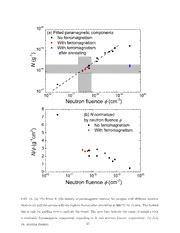
Defect induced magnetism in bulk SiC: interplay between ferromagnetism and paramagnetism Yutian Wang,1,2 Yu Liu,1,3 Elke Wendler,4 Rene Huebner,1 Wolfgang Anwand,5 5 1 Gang Wang,3 Xuliang Chen,6 Wei Tong,7 Zhaorong Yang,6 Frans Munnik,1 Gregor Bukalis,8 0 2 Xiaolong Chen,3 Sibylle Gemming,1,9,10 Manfred Helm,1,2,10 and Shengqiang Zhou1,∗ v o 1Helmholtz-Zentrum Dresden-Rossendorf, Institute of Ion Beam Physics N and Materials Research, Bautzner Landstr. 400, 01328 Dresden, Germany 0 1 2Technische Universit¨at Dresden, 01062 Dresden, Germany ] 3Research & Development Center for Functional Crystals, Beijing i c s National Laboratory for Condensed Matter Physics, Institute of - l r Physics, Chinese Academy of Sciences, Beijing 100190, China t m 4Institut fu¨r Festk¨orperphysik, Friedrich-Schiller Universit¨at Jena, 07743 Jena, Germany . t a m 5Helmholtz-Zentrum Dresden-Rossendorf, Institute of Radiation - Physics, Bautzner Landstr. 400, 01328 Dresden, Germany d n o 6Key Laboratory of Materials Physics, Institute of Solid State Physics, c [ Chinese Academy of Sciences, Hefei 230031, People’s Republic of China 2 7High Magnetic Field Laboratory, Hefei Institutes of Physical Science, v 6 Chinese Academy of Sciences, Hefei 230031, People’s Republic of China 9 0 8Helmholtz-Zentrum Berlin fu¨r Materialien und Energie GmbH, 1 0 Abteilung F-A1, Hahn-Meitner-Platz 1, 14109 Berlin, Germany . 1 0 9Faculty of Science, Technische Universit¨at Chemnitz, 09107 Chemnitz, Germany 5 1 10Center for Advancing Electronics Dresden, Technische : v i Universit¨at Dresden, 01314 Dresden, Germany X r (Dated: November 11, 2015) a 1 Abstract Defect-inducedferromagnetismhastriggeredalotofinvestigations andcontroversies. Themajor issue is that the induced ferromagnetic signal is so weak that it can sufficiently be accounted for by trace contamination. To resolve this issue, we studied the variation of the magnetic properties of SiC after neutron irradiation with fluence covering four orders of magnitude. A large param- agnetic component has been induced and scales up with defect concentration, which can be well accounted for by uncoupled divacancies. However, the ferromagnetic contribution is still weak and only appears in the low fluence range of neutrons or after annealing treatments. First-principles calculations hint towards a mutually exclusive role of the concentration of defects: a higher defect- concentration favors a larger magnetic interaction at the expense of spin polarization. Combining both experimental and first-principles calculation results, the defect-induced ferromagnetism can probably be understood as a local effect which cannot be scaled up with the volume. ∗ Electronic address: [email protected] 2 I. INTRODUCTION The magic of magnetism was disclosed in the early 20th century with the development of quantum mechanics. The Heisenberg model has since then been extremely successful to understand magnetism and magnetic materials [1]. All previously identified ferromagnetic bulk materials contain elements with partially filled 3d (4d) or 4f (5f) shells. A fundamen- tal question is whether materials containing only s or p electrons can be ferromagnetic. For nearly two decades, there have been various theoretical and experimental studies devoted to clarifying this question [2–20]. It turns out that materials with completely filled 3d or 4f shells or with only s or p electrons can be ferromagnetic when they contain defects. Among those materials, graphite/graphene and oxides attract particular attention due to experi- mental evidence reported by various groups [3, 5, 10, 21–27]. However, the experimentally measured ferromagnetism remains a weak signal slightly above the detection limit of sen- sitive SQUID magnetometry [9, 17, 28–31]. The very weak magnetization not only limits the practical applications, but also raises questions on the fundamentals of defect induced ferromagnetism. On the one hand, measurement artifacts in SQUID magnetometry may occur: improper mounting of samples and wrong use of sample holders can easily generate ferromagnetic like signal [32–34]. On the other hand, the debate over the purity of graphite and oxide substrates continues in parallel [35–39]. Pristine graphite and oxides substrates are often ferromagnetic due to different contaminations or due to intrinsic defects [36, 40– 42], which hamper the interpretation of the observed ferromagnetism. Indeed, Nair et al. reported that there is only paramagnetism in graphene after introducing adatoms or defects [43]. Therefore, the understanding of defect-induced ferromagnetism in materials without par- tially filled 3d or 4f electron shells is far from satisfactory. It is rather calling for an investigation in which the following requirements should be fulfilled: • The pristine materials should be well controlled with the highest purity grade possible. • The materials should be supplied in a large quantity with identical properties, such that one can perform a series of experiments using identical specimens. • The materials should be free of elements with 3d or 4f electrons. 3 • The induced effect should exist in a large volume to measure a large enough magnetic signal. The last point was firstly proposed by Coey et al., who suggested to measure a bulk graphite-nodule to gain a large ferromagnetic signal [2]. However, minor amounts of mag- netite, kamacite, etc, also appear in their graphite samples [2]. These secondary phases are responsible for about two-thirds of the observed magnetization and the remaining one-thirds is attributed to the graphite-nodule. After screening by these facts, Si and SiC are the best candidates. Defect-induced ferro- magnetism was revealed in SiC after neutron and ion irradiation [30, 31]. Recently, it was suggested that the p electrons from the carbon atoms are mainly responsible for the long- range ferromagnetic coupling in SiC after ion irradiation [44], which is similar to the obser- vation in graphite [45]. In this work, we start with 4H-SiC, high purity semi-insulating SiC. Neutron irradiation was used to introduce defects in the whole sample volume over a large range of defect concentrations. The large “magnetic volume” and well controlled pristine materials allow for reliable interpretation for the following experimental observations: (1) paramagnetism increases with neutron fluence and saturates at the end; (2) ferromagnetism only occurs in a narrow fluence range although it is weak; (3) thermal annealing param- agnetic defective SiC can induce weak ferromagnetism. Together with density-functional theory (DFT), we show that there is an intrinsic limit to increase the ferromagnetic contri- bution to the magnetization. II. EXPERIMENT Commercial semi-insulating 4H-SiC single crystal wafers (Cree) were used for our experi- ment. Particle induced X-ray emission (PIXE) was performed to check magnetic impurities in the pristine sample. The concentrations of magnetic impurities (Fe, Co and Ni) are proved to be below the detection limits (shown later). Neutron irradiation was performed at chambers DBVK and DBVR at the BER II reactor at Helmholtz-Zentrum Berlin at a tem- perature less than 50 ◦C. The fluxes of DBVK are 1.08×1014 cm−2s−1 of thermal neutrons, 7.04×1012 cm−2s−1 of epithermal neutrons, and 5.80×1013 cm−2s−1 of fast neutrons, respec- tively and those of DBVR are 1.50×1014 cm−2s−1, 6.80×1012 cm−2s−1, 5.10×1013 cm−2s−1, correspondingly. Comparing with epithermal or fast neutrons, thermal neutrons only pro- 4 duce negligible displacement. Therefore, only epithermal and fast neutrons are accounted for in the fluence calculation [46]. The neutron fluence spanned a largerange from3.28×1016 cm−2 to 3.50×1019 cm−2 covering all possibilities from light damage to near amorphization. It is worthy to note that the applied neutron fluence covers the range used in Ref. [30] and is extended to much higher fluence values by two orders of magnitude. For structural characterization, µ-Raman spectroscopy has been performed by using a Nd:YAG Laser with 532 nm wavelength in the scattering geometry using a liquid nitrogen cooled charge-coupled device camera. Positron annihilation Doppler broadening spectroscopy (DBS) was applied to clarify the nature of defects using the mono-energetic slow position beam ”SPONSOR” [47]. High-resolution transmission electron microscopy (HR-TEM) investigations were done with an image-corrected Titan 80-300 microscope (FEI) operated at an accelerating voltage of 300 keV. The magnetic properties are measured by a superconducting quantum interfer- ence device magnetic property measurement system and vibrating sample magnetometers (SQUID-MPMS and SOUID-VSM, Quantum Design). The electron spin resonance (ESR) spectroscopy was performed at 9.4 GHz using a Bruker spectrometer (Bruker ELEXSYS E500). III. RESULTS A. Virgin vs. irradiated SiC Beforewe start to describe the detailed results, we first present the key factsfor the virgin SiC substrates employed in our investigation. To check the trace presence of transition metals (Fe, Co and Ni, etc.) in our SiC sub- strates, we have performed PIXE using 2 MeV protons with a broad beam of around 1 mm2, as shown in Figure 1. PIXE is a sensitive method to detect trace impurities in bulk volume without significant structural destruction [35, 39]. In the spectrum, the sharp peak is the Si K-line X-ray emission. The broad bump is due to the secondary electron Bremsstrahlung background. Forcommerciallyavailable, purest graphite, thereisdetectabletransitionmetal contamination (mainly Ti, V, Fe, Ni) as revealed by PIXE [39]. In sharp contrast, the tran- sition metal impurity in the SiC we used for this study, if there is, is below the detection limit of around 1 ppm (Fig. 1). 5 4H-SiC, Cree ) 5 Si K-edge s10 t n u o c ( 4 y10 t i s n e FeCoNi t n 3 I10 2 10 1 2 3 4 5 6 7 8 9 10 Energy (keV) FIG. 1: PIXE spectrum for the 4H-SiC wafer by a broad proton beam. Within the detection limit, no Fe, Co or Ni contamination is observed. Figure 2 shows a comparison of the magnetic properties measured at 5 K between the virgin SiC and a specimen irradiated with neutrons with a relative low fluence value of 4.68×1017 cm−2. The results are presented without any background subtraction but only normalized to the mass of the samples. The difference between the virgin and irradiated SiC is obviously not trivial: the virgin SiC shows purely diamagnetic behavior, while the irradiated one shows a paramagnetic component as revealed by the large deviation from the linear dependence on the magnetic field. The inset of Fig. 2 shows the zoom of the low field part. Besides the paramagnetic component induced by neutron irradiation, a small ferromagnetic contribution also shows up with its saturation magnetization around 1% of theparamagneticsignal. Thedefect-inducedparamagnetismandferromagnetismareexactly the key facts we are going to discuss in this manuscript. 6 ) @ 5 K g 0.01 4H-SiC, virgin / 17 -2 u 4.68 10 cm m e ( 0.00 n o ) ati mu/g5 z -0.01 4 e i - 00 t 1 e ( n M -5 g a -0.02 -2000 -1000 0 1000 2000 M Field (Oe) -40000 -20000 0 20000 40000 60000 Field (Oe) FIG. 2: Comparison of the magnetic properties: virgin vs. neutron irradiated SiC. Inset: the zoom of the low field part. Neutron irradiation induces significant, unambiguous magnetic variation in SiC. B. Structural properties 1. Raman spectroscopy The damage to the crystallinity of SiC upon neutron irradiation is verified by Raman spectroscopy. Figure 3 exemplarily shows the Raman spectra for the virgin sample and the samples irradiated with different neutron fluence values as indicated in the figure. The typical Raman scattering modes for 4H-SiC are resolved: folded transverse optic (FTO) and longitudinal optic (FLO) modes [48]. Note the different scale in the y-axis: with increasing fluence, the intensity of the Raman scattering modes is dramatically reduced, as observed in Refs. [30, 31]. This behaviour directly reflects the increasing disorder of the crystalline material. For the sample with largest fluence, the Raman modes are much broad and hardly ◦ resolved due to the large amount of defects [46, 49]. However, after annealing at 900 C for 15 min the crystalline order can be substantially recovered. 7 1000 FTO FLO 500 Virgin 0 300 200 ) t 100 17 -2 i n 6.90 10 cm u 0 b. 150 r a ( 100 y 18 -2 t 1.48 10 cm si 50 n e 0 t n i 30 18 -2 n 6.75 10 cm a m 20 a 10 R 0 20 10 19 -2 3.50 10 cm 0 700 750 800 850 900 950 1000 -1 Raman shift (cm ) FIG. 3: Raman spectra for virgin and neutron irradiated 4H-SiC single crystals. The folded transverseoptic(FTO)andlongitudinaloptic(FLO)modesareidentified. Withincreasingneutron fluence the Raman scattering intensity is decreased and the peak is broadened. Figure 4 shows the Raman spectra of an irradiated SiC (fluence: 3.50×1019 cm−2) after annealing at different temperature. The annealing was performed in N atmosphere at 800, 2 ◦ 900 and 1000 C for 15 min. With increasing annealing temperature, the peaks of the SiC Raman modes become sharper and their intensity gradually increases. The increase of the peak intensity is related to the recovery of the crystalline lattice with annealing. However, a complete recovery of the lattice is not achieved. It has been shown that after annealing at 8 ) it 250 n u 19 -2 3.50 10 cm . b 200 r a ( y it 150 o s 1000 C n e o nt 100 900 C i o n 800 C a m 50 Non-annealed a R 0 700 750 800 850 900 950 1000 -1 Raman shift (cm ) FIG. 4: Raman spectra for neutron irradiated and thermally annealed 4H-SiC single crystals. The annealing was performed in N atmosphere 15 min at the temperature indicated. With increasing 2 annealing temperature, the structural damage has been healed. ◦ 1450 C for 10 min the Raman peaks are comparable to the pristine SiC [50]. 2. Positron annihilation spectroscopy Positron annihilation Doppler broadening spectroscopy (DBS) is an excellent technique to detect open volume defects from clusters consisting of several vacancies down to a mono- vacancy [47]. The positron in a crystal lattice is strongly subjected to repulsion from the positive atom core. Because of the locally reduced atomic density inside the open volume defects, positrons have a high probability to be trapped and to annihilate with electrons in these defects by the emission of two 511 keV photons. The Doppler broadening of the 511 keV annihilation line is mainly caused by the momentum of the electron due to the very low momentum of the thermalized positron. The Doppler broadening parameter S, obtained from the 511 keV annihilation line, reflects the fraction of positrons, annihilating with electrons of low momentum (valance electrons). In this study, the S parameter is 9 defined as the ratio of the counts from the central part of the annihilation peak (here 510.17 keV - 511.83 keV) to the total number of counts in the whole peak (498 keV - 524 keV). Therefore, the S parameter is a measure for the open volume in the material. It increases with increasing size of the particular open volume defects. According to our previous positron annihilation experiments [30, 31], divacancies have been identified in neutron or ion irradiated SiC. Figure 5(a) shows the measured S parame- ters versus the incident positron energy. The plateau of the S parameters of the irradiated samples above a positron energy of 2 keV corresponds to positron annihilation in the defects created by neutron irradiation. Defects are homogenously distributed along the depth as expected for neutron irradiation. The S parameters of irradiated samples are larger than that of the virgin SiC. S does not increase significantly with increasing neutron fluence, but does increase after annealing. Figure 5(b) displays the S − W plot for different sam- ples. A straight line is obtained in the S −W representation for different SiC samples. It shows that the same type of defects, i.e., open volume damage [51, 52], exist in different SiC samples. A relation between the ratio S /S and the number of agglomerated V V defect bulk Si C divacancies in 6H-SiC is published as a scaling curve in Ref. 51. As a rough estimation, the ◦ post-irradiation annealing at 900 C led to a defect agglomeration up to a size of 8 V V Si C divacancies. One has to note that PAS is only sensitive to negatively charged or neutral open volume defects. The S parameter increases with the size of the open volume defects, but not necessarily with the amount of defects. Raman and magnetization are measurements of total amount of defects. This explains why the S parameter increases after annealing, indicating coalescence of voids to larger but fewer ones, but the Raman and magnetization measurements show the decrease of amount of defects. 3. High-resolution transmission electron microscopy With the aim to visualize the defects, we performed high resolution transmission electron microscopy on selected samples: a pristine 4H-SiC, 431-56 (irradiated with neutron at the fluence of 3.50×1019 cm−2) and 431-56 after annealing at 900 ◦C for 15 min. Figure 6 dis- plays corresponding cross-sectional high-resolution transmission electron micrographs taken in [100] zone axis geometry. It is observed that the 4H-SiC stacking sequence (abcb) perpen- 10
