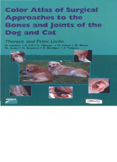
Color Atlas of Surgical Approaches to the Bones and Joints of the Dog and Cat. Thoracic and Pelvic Limbs PDF
Preview Color Atlas of Surgical Approaches to the Bones and Joints of the Dog and Cat. Thoracic and Pelvic Limbs
Color Atlas of Surgical .lpproaches t o the Thoracic and Pelvic Limbs R. Latorre I?. GI1 S. (3iment 0. Upez R. Henry M. Ayala G. Ramirez Fw Martinez V-ez Jw COLOR ATLAS OF SURGICAL APPROACHES TO THE BONES AND JOINTS OF THE DOG AND CAT Thoracic and pelvic limbs R. Latorre F. Gil S. Climent 0. L6pez R. Henry M. Ayala G. Ranlirez F. Martinez J. Viizquez XXI - 2009 Buenos Aires - Repljblica Argentina All rights reserved. No part of this publication may be reproduced, stored in a retrieval system, or transmitted, in any form or by any means, elec- tronic, mechanical, photocopying, recording, or otherwise, without prior written permission from Editorial Inter-Medica S.A. Deposit was made under the law 11.723 ISBN: 978-950-555-347-1 0 2009 b yE ditorial Inter-Medica S.A.I.C.I. Junin917Piso lo"A"-C1113AAC Ciudad Autonoma de Buenos Aires - Republics Argentina Tels.: (54-1 1) 4961-7249 14961-9234 14962-3145 FAX: (54-1 1) 4961-5572 E-mail: [email protected] E-mail: ventasBinter-medica.com.ar http://www.inter-medica. com.ar ww.seleccionesveterinarias.com Latorre, Rafael Color atlas of surgical approaches to the bones and joints of the dog and cat: toracic and pelvic. - la ed. Buenos Aires: Inter-Medica. 2009. 272 p.; 28x20 cm. I I ISBN 978-950-555-347-1 I 1. Veterinary medicine. 2. Surgery. I. Tittle CDD 636.089 Print in Talleres Graficos Valdez Loyola 1568 - Buenos Aires Impreso en Argentina - Printed in Argentina Tirada: 5000 ejemplares Este libro se termino de imprimir en Enero de 7009. leans. elec- Autores colaboradores ARENCIBIEAS PINOSAA, . MARTINEZG OMARIZF,. DVM, PhD. Professor of Veterinary Anatomy, DVM, PhD. Professor of Veterinary Anatomy, University University of Las Palmas de Gran Canaria, Spain. of Murcia, Spain. AYALAF LORENCIANMO,a .D . ORENESH ERNANDEZM, . DVM, PhD. Professor of Veterinary Anatomy, University Technical Specialist of Veterinary Anatomy, University of Murcia, Spain. of Murcia, Spain. ALBARRACL~ON PEZ,J . RAM~REZZAR ZOSA G. Auxiliary Technical Specialist of Veterinary Anatomy, DVM, PhD. Professor of Veterinary Anatomy, University University of Murcia, Spain. of Murcia, Spain. CLIMENTP ERIS,S . ROJOR ios, D. DVM, PhD. Professor of Veterinary Anatomy, University DVM. Professor of Veterinary Anatomy, University of of Zaragoza, Spain. Murcia, Spain. CLIMENTA ROZ,M . Ros SEMPEREJ, . DVM. Professor of Veterinary Anatomy, University of DVM. . Professor of Veterinary Anatomy, University of Zaragoza, Spain. Murcia, Spain. DRAPE,J . RUIZ,M . DVM, PhD. Aquivet Veterinary Hospital Director, DVM, PhD. Director of the Mediterranean Veterinary Eysines, Burdeoux, France. Hospital, Madrid, Spain. GILC ANO,E SANCHEMZ ARGALLOF,. DVM, PhD. Professor of Veterinary Anatomy, University DVM, PhD. Scientific Director of Minimally lnvasive of Murcia, Spain. Surgery Center Jesus Uson, Caceres, Spain. HENRY,R . SANCHEZC OLLADOC, . DVM, PhD. Professor of Veterinary Anatomy, University DVM. Professor of Veterinary Anatomy, University of of Tennessee, USA. Murcia, Spain. KOSTLIN,R . USONG ARGALLOJ,. DVM, PhD. Professor of Veterinary Surgery, University DVM, PhD. Director of the Foundation Minimally of Munich, Germany. lnvasive Surgery Center Jesus Uson, Caceres, Spain. LATORRER EVIRIEGOR, . V~QUEAZU TON,J . DVM, PhD. Professor of Veterinary Anatomy, University DVM, PhD. Professor of Veterinary Anatomy, University of Murcia, Spain. of Murcia, Spain. LOPEZA LBORS,0 . VEREZF RAGUELAJ,. L. DVM, PhD. Professor of Veterinary Anatomy, University DVM, PhD. Director of the Veterinary Hospital of Murcia, Spain. Ultramar Clinic, El Ferrol, Spain. LOSILLAG UUAS,S . ZAERA, J.P. DVM. Endoluminal Therapy and Diagnosis Unit, DVM, PhD. Professor of Veterinary Surgery, University Minimally lnvasive Surgery Center Jesus Uson, of Las Palmas de Gran Canaria, Spain. Caceres, Spain. .. . lll Preface Many surgeons usually choose to review regional anatomy when planning for surgery. Anatomical review is more likely while learning new surgical techniques, as identification of anatomical structures is not as routine. This atlas provides an answer to traumatologists who have been asking for a collection of colour anatomical images of the most common surgical approaches to the limbs. The selected images have been used in continuing education courses for traumatologists with great success and availability in text book format is often asked. The approaches to the thoracic limb are presented in three sections. The first includes the scapula, shoulder and hume- . . rus of the dog, the second contains the elbow, radius, ulna and manus of the dog, and a third section includes selections on the cat. The pelvic limb begins with the hip joint and thigh and continues with the knee, leg and pes of the dog. It conclu- des with the corresponding approaches in the cat. Images of the articulated bones of the region are presented at the begin- ning of each section. All approaches were completed on fresh tissue (no fixation) for more natural colour. Cadaver vessels were highlighted by colour injection. Superficial to deep views of preparations are presented with the relevant muscles, liga- ments, nerves and vessels identified. Additionally, sevcral videos of the thoracic and pelvic limbs with 3D reconstruction, obtained from live specimens at the Minimally Inuasiw Surgery CentreJesus Usdn (Ciceres, Spain) with a "BV Pulsera 3D- RX Option. Philips. S. A." device, are included. Indications for each approach are referenced at the beginning of each chap- ter. All approaches were carried out on left limbs - with the exception of some in the manus and pes- , and sequenced from proximal to distal. Footnotes indicate the commonly used protocol for each surgical approach. We would like to conclude with a very special reference to Prof Dr. Francisco Moreno Medina, who had to leave his care- er in anatomy early and retire due to illness. He founded the Anatomy and Embryology group at the University of Murcia and from him we inherited a large part of our anatomical knowledge and passion for working in the dissection room. THE AUTHORS "Anatomy without clinic is dead, clinics without anatomy is deady" (Platzer) (None of the specimens was eutharratized for dissection purposes. All cadavers were obtainedfrom the Animal Facility of the University ofMurcia, which oversees nllprotocols for Animal Healthcare and is accredited by the European Bureau for protection of research risks in animals. Most cahuers wereper+sed with coloured chemicalsf or a better identification of arteries and veins). I Section I Mediopalmar approach to the carpal joint . . . . . 95 . . . . . . . . . . . . . Dog, thoracic limb I Approach to the metacarpal bones. . . . . . . . . . . . 99 Approach to the phalanges and the Scapula, shoulder joint and humerus interphulungeal joints . . . . . . . . . . . . . . . . . . . 103 Anatomical considerations . . . . . . . . . . . . . . . . . . . . 3 Approach to the lateral suface, spine Section 2 . . . . . . . . . . . . . and acromion of the scapula . . . . . . . . . . . . . . 9 Cat, thoracic limb 107 Craniolaterul approach to the shoulder hint hv ~rrnmianl stentnm~~ 1 3 Anatomical considerations . . . . . . . . . . . . . . . . . . . . 108 . . . . . . . . . . . . . . . . Caudolateral approach to the shoulder joint . . . . 2 1 Humerus: approach to the distalportion of the Craniomedial approach to the shoulder joint . . . . 25 diaphysis by craniol ateral incision . . . . . . . . . . 1 19 Approach to the proximal diaphysis of the humerus 3 1 Approach to the distal humeral diaphysis " and the humeral supracondylar region - -roach to the medial humeral diaphysis via a via a medial incision . . . . . . . . . . . . . . . . . . . 123 3 c medial incision . . . . . . . . . . . . . . . . . . . . . . . 47 Section 3 . . . . . . . . . . . . . . . Elbow, ulna and manus Dog, pelvic limb 139 Anatomical considerations . . . . . . . . . . . . . . . . . . . . 5 1 The pelvis and hip (coxal) joint Lateral approach to the humeral condyle Anatomical considerations . . . . . . . . . . . . . . . . . . . . 14 1 Approach to the humeroulnarpart of the elbow joint Approach to the ventral surface of the sacrum . , . 155 medial aspect of the humeral condyle via Approach to the craniodorsal and caudodorsal intermuscular incision . . . . . . . . . . . . . . . . . . 67 the trochlear notch . . . . . . . . . . . . . . . . . . . . . 7 1 Approach to the caudodorsal regions of Approach to the olecranon tuber . . . . . . . . . . . . . 73 the hip joint with gluteal muscle tenotomy . . . . 165 Approach to the distal ulnar diaphysis and Approach to the os coxae . . . . . . . . . . . . . . . . . . 167 and diaphysis ofthe radius . -. . . . . . . . . . . . . . . 79 Approach to the pubis and the pelvic symphysis . . 175 Approach to the diaphysis of the radius via a Approach to the ischium . . . . . . . . . . . . . . . . . . 179 . . I I . eazal zncznon . . . . . . . . . . . . . . . . . . . . . . . 81 Approach to the diaphysis of the femur . . . . . . . . 1 87 Dorsal approach to the carpal joint . . . . . . . . . . 9 1 Contents Stifle. leg and foot Approach to the calcaneus and the plantar surface Anatomical considerations .................... 189 ofthe tarsal bones ..................... 237 I Approach to tile distalfemur and stzfle joint Section 4 I via a lateral incision ................... 20 1 Cat. pelvic limb ................ 239 Approach to the medial collateral ligament and the caudomedial region of the sttjle joint .... 205 Anatomical considerations .................... 240 Approach to the lateral collateral ligament of the caudolateral stzjle joint region ............ 209 Approach to the wing of the ilium by lateral incision ...................... 247 Approach to the proximal tibia via a medial incision ...................... 21 3 Craniodorsal and caudodorsal approaches to the hip joint by osteotomy of the major ti-ochanter .... 251 Approach to the tibial diaphysis ............. 2 19 Approach to the dyaphisis of the femur ........ 255 Approach to the lateral malleolus and tarsocruraljoint ................... 223 Approach to the stzjle joint by luteral incision ... 259 Approach to the medial malleolus Approach to the dy~phisiso f the tibia ......... 263 and tarsocruralj oint ................... 227 Approach to the tarsocruralj oint via osteotomy References .............................. 266 of the medial malleolus .................. 23 1 Approach to the calcaneus ................. 235 Thoracic limb Chapter 1 Skeleton of dog, left view Scapula, shoulder joint and humerus Anatomical considerations mara rk L
Description: