Atlas of Human Anatomy and Surgery. Nevrologia PDF
Preview Atlas of Human Anatomy and Surgery. Nevrologia
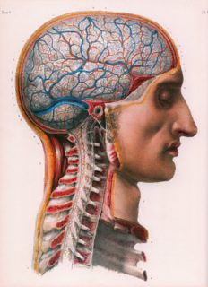
Yomme 3 N V R O LO G IA : e S N C , y s t e m a e r v o s u m e n t r a l e P , A . e r i p h e r i c u m e t u t o n o m i c u m O S r g a n a e n s u u m N e u r o l o g y : C e n t r a l , p e r i p h e r a l , and v e g e t a t i v e nervous s y s t e m S e n s o r y organs N ÉVROLOGIE: S y s t è m e n e r v e u x c e n t r a l , PÉRIPHÉRIQUE, ET AUTONOME. O rga nes des s en s N e u r o l o g i e : Ze n t r a l e s , p e r i p h e r e s UND VEGETATIVES NERVENSYSTEM. S i n n e s o r g a n e < VOLUME 3. PLATE 8. < TOME 3. PLANCHE 8. MENINGES MÉNINGES Right lateral view. Vue latérale droite. Dura mater removed, showing the arachnoid and Dure-mère enlevée démontrant l’arachnoïde et the pia mater and the superficial vessels. la pie-mère et les vaisseaux superficiels. vo Lu m e 3. piaT es 1 ,2 ,3 * MENINGES 1 - MENINGES 1 - MENINGES 1 - MENINGEN Overview of the central nervous system. Anterior view (after the vertebral Ensemble du système nerveux central. Vue antérieure (après ouverture du Cesamtes Zentralnervensystem. Vorderansicht (nach Eröffnung des canal and the skull have been opened). On the subject’s right half, the dura canal vertébral et du crâne). Sur la moitié droite du sujet, dure-mère en Wirbelkanals und des Schädels). Rechte Körperseite: Dura mater belas mater is in place. On the left half, the dura mater has been opened, show place. Sur la moitié gauche, dure-mère ouverte, démontrant l’arachnoïde sen. Linke Körperseite: Dura mater zur Darstellung von Arachnoidea und ing the arachnoid and the pia mater. et la pie-mère. Pia mater eröffnet. MENINGES MENINGES 2-MENINGEN % — % — Overview of the central nervous system. Posterior view (after the vertebral Ensemble du système nerveux central. Vue postérieure (après ouverture Gesamtes Zentralnervensystem. Rückansicht (nach Eröffnung des Wirbel canal and the skull have been opened). On the subject’s left half, the dura du canal vertébral et du crâne). Sur la moitié gauche du sujet, dure-mère kanals und des Schädels). Linke Körperseite: Dura mater belassen. Rechte mater is in place. On the right half, the dura mater has been opened, show en place. Sur la moitié droite, dure-mère ouverte, démontrant l’arachnoï Körperseite: Dura mater zur Darstellung von Arachnoidea und Pia mater ing the arachnoid and the pia mater. de et la pie-mère. * eröffnet. 3-MENINGES 3-MÉNINGES 3-MENINGEN Overview of the central nervous system. Right lateral view (after paramedi Ensemble du système nerveux central. Vue latérale droite (après section Gesamtes Zentralnervensystem. Ansicht von rechts-lateral (Sagittalpara- an sagittal section of the spine and opening of the skull). Dura mater in sagittale paramédiane droite de la colonne vertébrale et ouverture du medianschnitt rechts der Wirbelsäule. Schädel eröffnet). Dura mater be place. crâne). Dure-mère en place. lassen. *94 voLume 3. PLare 4. MENINGES ENCEPHALI MENINGES OF THE BRAIN OR CRANIAL MÉNINGES DE L’ENCÉPHALE MENINGEN DES SCHÄDELS ODER MENINGES OU CRÂNIENNES HIRNHÄUTE Superior view of the brain. On the subject’s left half the dura mater is in Vue supérieure du cerveau. Sur la moitié gauche du sujet, dure-mère en Ansicht des Großhirns von oben. Linke Körperseite: Dura mater belassen. place. On the right half the dura mater is opened, showing the arachnoid place. Sur la moitié droite, dure-mère ouverte démontrant l'arachnoïde et Rechte Körperseite: Dura mater zur Darstellung von Arachnoidea und Pia and the pia mater. On the median line, the superior sagittal venous sinus la pie-mère. Surla ligne médiane, lesinus veineux sagittal supérieurest en mater eröffnet. Auf der Medianlinie ist der Sinus sagitlalis superior teil has been partly opened. partie ouvert. weise freipräpariert. 196 voLume 3. PLare 5 MENINGES ENCEPHALI * ' MENINGES OF THE BRAIN OR CRANIAL MÉNINGES DE L’ENCÉPHALE MENINGEN DES SCHÄDELS ODER MENINGES OU CRÂNIENNES HIRNHÄUTE Inferior view of the brain. On the subject's right half the dura mater is in Vue inférieure de l'encéphale. Sur la moitié droite du sujet, dure-mère en Ansicht des Schädels von unten. Rechte Körperseite: Dura mater belassen. place. On the left half the dura mater is opened, showing the arachnoid and place. Sur la moitié gauche, dure-mère ouverte démontrant l’arachnoïde et Linke Körperseite: Dura mater zur Darstellung von Arachnoidea und Pia the pia mater. la pie-mère. mater eröffnet. >97 vomme 3. PLclTe 6, 7, 9, 10 * MENINGES ENCEPHALE MENINGES SPINALES 6 - MENINGES OF THE BRAIN OR CRANIAL 6 - MÉNINGES DE L’ENCÉPHALE 6 - MENINGEN DES SCHÄDELS ODER MENINGES OU CRÂNIENNES HIRNHÄUTE Superior views. Vues supérieures. Ansichten von oben. Fig. i. - Falx cerebri maintained, with the superior sagittal venous sinus Fig. i. - Faux du cerveau conservée avec le sinus veineux sagittal supérieur Abb. i. - Großhimsichel mit eröffnetem Sinus sagittalis superior erhalten. opened. Tentorium cerebelli in place, with the incisura tentorii. Anterior ouvert. Tente du cervelet en place avec l’incisure de la tente. Fosses crâ Kleinhimzelt mitTentoriumschlitz belassen. Vordere und mittlere Schädel and middle cranial fossae. niennes antérieure et moyenne. grube. Fig. z. - Falx cerebri and left half of the tentorium cerebelli removed. Fig. 3. - Faux du cerveau et moitié gauche de la tente du cervelet enlevées. Abb. a. - Großhimsichel und linke Hälfte des Kleinhimzelts entfernt. Venous sinuses of the dura mater opened. Anterior, middle, and posterior Sinus veineux de la dure-mère ouverts. Fosses crâniennes antérieure, Venöser Sinus der Dura mater eröffnet. Vordere, mittlere und hintere cranial fossae. moyenne, et postérieure. Schädelgrube. 7 - MENINGES OF THE BRAIN OR CRANIAL 7 - MÉNINGES DE L’ENCÉPHALE 7 - MENINGEN DES SCHÄDELS ODER MENINGES OU CRÂNIENNES HIRNHÄUTE Fig. l. - Antero-superior view. Falx cerebri removed over the best part of its Fig. î. - Vue antéro-supérieure. Faux du cerveau enlevée sur l’essentiel de Abb. i. - Vorderansicht von schräg oben. Großhirnsichel, wesentliche length. Tentorium cerebelli in place, with the incisura tentorii. Anterior son étendue. Tente du cervelet en place avec l’incisure de la tente. Fosses Teile entfernt. Kleinhimzelt mit Tentoriumschlitz belassen. Vordere und and middle cranial fossae. crâniennes antérieure et moyenne. mittlere Schädelgrube. Fig. 3. - Oblique right supero lateral view. Falx cerebri and tentorium cere Fig. 3. - Vue oblique supérieure latérale droite. Faux du cerveau et tente du Abb. a. - Ansicht von schräg oben rechts. Großhirnsichel und Kleinhirn belli in place, with the incisura tentorii. cervelet en place avec l’incisure de la tente. zelt mitTentoriumschlitz belassen. 9-MENINGES 9-MENINGES 9-MENINGEN Fig. i. - Overview of the central nervous system. Demonstration of the sub Fig. 1. - Ensemble du système nerveux central. Démonstration des espaces Abb. 1. - Gesamtes Zentralnervensystem. Darstellung der mit Liquor ge arachnoid spaces and cisterns which are filled with the cerebrospinal et citernes subarachnoïdiens occupés par le liquide cérébrospinal. Coupe füllten Subarachnoidalräume und Zisternen. Sagittalmedianschnitt, An fluid. Median sagittal section. Right lateral view of the left half. sagittale médiane, vue latérale droite de la moitié gauche. sicht der linken Körperseite von rechts-lateral. Fig. a. - Arrangement at the level of the first cervical vertebra (atlas). Super Fig. 3. - Disposition au niveau de la première vertèbre cervicale (atlas). Abb. *. - Anordnung auf Höhe des ersten Halswirbels (Atlas). Ansicht von ior view. Vue supérieure. oben. 3 3 3 Fig. . - Arrangement at the level of a thoracic vertebra. Axial section. Fig. . - Disposition au niveau d'une vertèbre thoracique. Coupe axiale. Abb. . - Anordnung auf Höhe eines Brustwirbels. Axialschnitt. Fig. 4. - Arrangement at the level of the first lumbar vertebra. Axial section. Fig. 4. - Disposition au niveau de la première vertèbre lombaire. Coupe Abb. 4. - Anordnung auf Höhe des ersten Lendenwirbels. Axialschnitt. Fig. 5. - Arrangement at the level of the fifth lumbar vertebra. Axial section. axiale. Abb. 5. - Anordnung auf Höhe des fünften Lendenwirbels. Axialschnitt. Fig. 5. - Disposition au niveau de la cinquième vertèbre lombaire. Vue axiale. 10-SPINAL MENINGES 10 - MÉNINGES SPINALES 10 - RÜCKENMARKHÄUTE Fig. 1. - Anterior view. Upper two-thirds of the spinal cord. Dura mater Fig. 1. - Vue antérieure. Deux-tiers supérieurs de la moelle spinale. Dure- Abb. 1. - Vorderansicht. Obere zwei Drittel des Rückenmarks. Dura mater opened, includes the denticulate ligament. mère ouverte avec le ligament dentelé. mit Ligamentum denticulatum eröffnet. Fig. a. - Anterior view. Lower third of the spinal cord with the conus Fig. 3. - Vue antérieure. Tiers inférieur de la moelle spinale avec le cône Abb. 3. - Vorderansicht. Unteres Drittel des Rückenmarks mit Conus medullaris and the cauda equina. Dura mater opened, includes the den médullaire et la queue de cheval. Dure-mère ouverte avec le ligament den medullaris und Cauda equina. Dura mater mit Ligamentum denticulatum ticulate ligament. telé. eröffnet. 3 3 3 Fig. . - Posterior view. Upper two-thirds of the spinal cord. Dura mater Fig. . - Vue postérieure. Deux-tiers supérieurs de la moelle spinale. Dure- Abb. . - Rückansicht. Obere zwei Drittel des Rückenmarks. Dura mater opened, includes the denticulate ligament. mère ouverte avec le ligament dentelé. f mit Ligamentum denticulatum eröffnet. Fig. 4. - Posterior view. Lower third of the spinal cord with the conus Fig. 4. - Vue postérieure. Tiers inférieur de la moelle spinale avec le cône Abb. 4. - Rückansicht. Unteres Drittel des Rückenmarks mit Conus medullaris and the cauda equina. Dura mater opened, includes the den médullaire et la queue de cheval. Dure-mère ouverte avec le ligament den medullaris und Cauda equina. Dura mater mit Ligamentum denticulatum ticulate ligament. telé. eröffnet. Fig. 5. - Filum terminale between the lower end of the spinal cord, or conus Fig. 5. - Filament terminal entre l’extrémité inférieure de la moelle, ou Abb. 5. - Filum terminale zwischen äußerstem Ende des Rückenmarks medullaris, and the posterior aspect of the coccyx. cône médullaire, et la face postérieure du coccyx. bzw. Conus medullaris und der Rückseite des Steißbeins. Fig. 6. - Detail of an attachment of the denticulate ligament. Fig. 6. - Détail d'une attache du ligament dentelé. Abb. 6. - Detail einer Ansatzstelle des Ligamentum denticulatum. Fig. 7. - Arrangement at the level of the first cervical vertebra (atlas). Super Fig. 7. - Disposition au niveau de la première vertèbre cervicale (atlas). Abb. 7. - Anordnung auf Höhe des ersten Halswirbels (Atlas). Ansicht von ior view. Vue supérieure. oben. Fig. 8. - Arrangement at the level of a thoracic vertebra. Axial section. Fig. 8. - Disposition au niveau d’une vertèbre thoracique. Coupe axiale. Abb. 8. - Anordnung auf Höhe eines Brustwirbels. Axialschnitt. Fig. 9. - Arrangement at the level of a lumbar vertebra. Axial section. Fig. 9. - Disposition au niveau d’une vertèbre lombaire. Coupe axiale. Abb. 9. -Anordnung auf Höhe eines Lendenwirbels. Axialschnitt. 198 voLume 3. PLare 15 ENCEPHALON ET CEREBRUM ENCEPHALON ENCÉPHALE GEHIRN Superior view in situ. Vue supérieure in situ. Ansicht von oben in situ. Right and left cerebral hemispheres. Hémisphères cérébraux droit et gauche. Rechte und linke Großhirnhemisphäre. 200 voLu m e 3. PLare i6 ENCEPHALON ET CEREBRUM ENCEPHALON ENCÉPHALE GEHIRN Inferiorviewinsitu. Vue inférieure in situ. Ansicht von unten in situ. Brain, cerebellum, and brainstem. Cerveau, cervelet, et tronc cérébral. Großhirn. Kleinhirn und Hirnstamm.
Description: